First Serv. O! I am slain. My lord, you have one eye left
To see some mischief on him. O! [Dies.]
Corn. Lest it see more, prevent it. Out, vile jelly! Where is thy lustre now?
King Lear
Act III. Scene VII.
It was one of those days. Sunny, a pleasant temperature, and the birds were singing. Just like 9/11. I had an appointment at the UM’s Kellog Eye Center. A stock intro, you say. Let me try another. The least painful procedure that day was the three novocain injections in my right eyeball near the end of my appointment.
Having undergone cataract surgery 3 years ago, I was convinced a plaque had once again built up on my right eye. This is not unusual, and requires a relatively simple procedure whereby the plaque is simply lasered away from the artificial lens. It wasn’t that.
The diagnostic required to know this can cause varying degrees of pain. Everyone knows dentistry can require sadistic procedures, but not as many are aware that so too can eye exams.
“IS IT SAFE?”
Per usual, the dilation drops were applied and the doc pulled up a blinding light and began to inspect the poor orb. The patient’s task is to systematically scan the perimeter of their vision as the eye is examined. The next step is to administer numbing drops topically and probe the eyeball in the socket with a cotton swab. Oh boy.
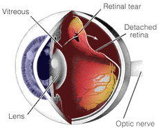
The vitreous humor -- the equivalent of the fluidic mass within the grape’s skin -- is gelatinous, and within limits, can be squeezed without bursting the soft tissue of the outer eyeball. Probing and manipulating the eyeball allows a close inspection of the back of the eye – the retina. What they were looking for were tears in the retina.
What it feels like is a scraping o f the eye with spoon or butter knife. The strain of fixing one’s gaze on a particular point in the room while the eye is squeezed and perused is both tiring and painful. “Do you need a break?” “No.”
Imagine the terra cotta color of the desert as a background. Then think of dried cracks in that parched landscape. Arid arroyos with burnt orange beds. Very surrealistic, an abstract landscape of pain, all so Daliesque. “That’s what  it looks like, doc.”
This wasn’t the Retina Clinic (third floor), mind you. Not yet. The doc sees something, but he can’t be sure, so he brings in a bigger doc. Funny how the big docs relegate the torturous procedures to their underlings. The big doc affixes an adjustable lens that holds the eye open while he dials in a macroscopic view. From a patient’s perspective it looks like those telescopic shots of the sun experiencing solar storms. Wow!
He confirms the medium doc’s conclusion that I should be seen in the Retina Clinic. So it’s up the elevator I go. The exam is the same except for an excruciating twist, my eye is held open with metal retractors ala A Clockwork Orange. I ask him about the dry riverbed frescoes I’m seeing. “Oh, you’re seeing the blood vessels that cover the outer eyeball. Nice.
My over-stressed, rigid, contorted body and face are now ready for the big doc. I know he’ll be gentle. This guys wearing a suit sans a jacket, and I immediately recognize him from the banners hanging in the atrium of this brand spanking new facility. He’s world famous (don’t we all say this about our doctors? It’s comforting). Like the first big doc, he attaches the super lens and looks long and hard. He announces that he’s found some suspect weak spots (potential tears), and that cryogenic ablation (cryopexy) is required. I ask when this might be scheduled, and he says, “right now, we have the cropexy room prepared.”
UN CHIEN ANDALOU
To return from whence I started, the preliminary to the website description that follows was the administration of three novocain shots to the eyeball.
Usually, retinal cryopexy is administered under local anesthesia. The procedure involves placing a metal probe against the eye. When a foot pedal is depressed, the tip of the cryopexy probe becomes very cold as a result of the rapid expansion of very cold gases (usually nitrous oxide) within the probe tip. When the probe is placed on the eye the formation of water crystals followed by rapid thawing results in tissue destruction. This is followed by healing and scar tissue formation.
In the case of retinal detachment, treatment calls for irritating the tissue around each of the retinal tears. Cryopexy stimulates scar formation, sealing the edges of the tear. This is typically done by looking into the eye using the indirect ophthalmoscope while pushing gently on the outside of the eye using the cryopexy probe, producing a small area of freezing that involves the retina and the tissues immediately underneath it. Using multiple small freezes like this, each of the tears is surrounded. Irritated tissue forms a scar, which brings the retina back into contact with the tissue underneath it. 
How did it feel? Ever had an ice cream headache, or what I call “brain freeze?” Each time the tip was applied to my eyeball it was like a laser brain freeze that extended from the eye deep into my brain. After multiple applications, I stood up, reeling from the chair to the door. Teary-eyed and feeling like Frazier after round 14 in his Manila fight with Ali, I’m told I can leave and they’ll see me Friday.
Best - Randy


